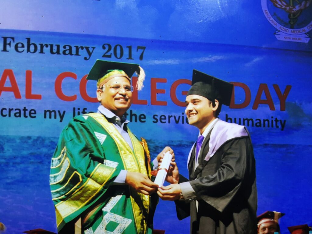
MAMC, Sports Injury Centre Safdarjung & Max Saket Trained
MBBS (MAMC, Delhi) · MS Ortho (MAMC, Delhi)
DNB Ortho · MNAMS · FIJR · FIASM · Dip Sports Med
Former Assistant Professor
Jamia Hamdard & HAHC Hospital, Delhi
Arthroscopy and Joint Replacement
Knee, Hip & Shoulder Specialist
Dashvanth Healthcare
Meet your specialist
Dr Shahrukh Khan
Consultant Orthopedic Surgeon
Arthroscopy and Joint Replacement
Knee, Hip & Shoulder Specialist
MBBS · MS Orthopaedics · DNB · MNAMS
MAMC, Sports Injury Centre Safdarjung & Max Saket Trained
Orthopaedic care shaped by training at Maulana Azad Medical College, the Sports Injury Centre at Safdarjung Hospital, and fellowship training in arthroscopy and joint replacement.
His practice brings together joint replacement, arthroscopy, sports injury care and fracture treatment, with clear explanations to help patients understand their options.
Clinical & academic journey
Experience that shapes his practice.
Senior Resident · Orthopaedics
Lok Nayak Hospital, New Delhi
Department of Orthopaedics.
Senior Resident
Sushruta Trauma Centre, Delhi
Trauma care.
Senior Resident
Sports Injury Centre, Safdarjung Hospital
Sports injuries and arthroscopy.
Assistant Professor
Jamia Hamdard & HAHC Hospital, Delhi
Orthopaedics — clinical and academic appointment.
Consultant
Max Smart Hospital, Saket, Delhi
Robotic joint replacement and arthroscopy · Dr SKS Marya’s team.
Current clinical practice · Director
Dashvanth Healthcare
Dr Shahrukh’s Bone & Joint Clinic, Krishna Nagar Extension, Delhi.
Current clinical practice · Consultant Orthopedic Surgeon
Apollo Spectra Hospitals, Delhi
Karol Bagh, New Delhi.
Beyond the consultation
Professional Journey & Training
Photographs from Dr Shahrukh Khan’s training and professional encounters.
Dr Shahrukh Khan at GSL-ABC Advanced Bio-Skills Centre, pictured with robotic surgical equipment.
A photograph from Dr Shahrukh Khan’s professional journey at Max Healthcare.
A certificate presentation photograph with Dr IPS Oberoi at Artemis Hospitals.
A moment from Dr Shahrukh Khan’s professional journey in orthopaedics.
With the Director of the Sports Injury Centre, Safdarjung Hospital.
Personal moments
Memorable encounters
With Mary Kom, six-time world boxing champion.
X-rays from Dr Shahrukh’s practice
X-ray & treatment gallery
Images supplied by Dr Shahrukh Khan, with short explanations of the bones and implants shown. Tap an image to enlarge it.
78 images · Choose a body area to explore
Knee15 images
X-ray showing a plate and screws in the upper shin bone near the knee.
X-ray showing wires and pins around the kneecap.
X-ray showing a plate and screws in the upper shin bone near the knee.
Views of the knee joint and surrounding bones.
Knee X-ray showing replacement components at the ends of the thigh and shin bones.
Views of the knee joint and surrounding bones.
X-ray showing knee replacement components with stems extending into the thigh and shin bones.
Knee X-ray views showing replacement components on one side of the joint.
Knee X-ray showing small fixation devices and skin staples.
X-ray showing a plate and screws in the lower thigh bone near the knee.
X-ray showing a plate and screws in the upper shin bone near the knee.
Knee X-ray showing replacement components at the ends of the thigh and shin bones.
Hip and pelvis13 images
An image taken during hip surgery, showing a hip implant and a surgical instrument.
Pelvic X-ray showing a plate, screws and a long transverse screw.
Hip and upper thigh X-ray showing a nail inside the bone with fixation screws.
X-ray of the hip joint and upper thigh bone.
An image of the hip area showing a guidewire or procedural instrument.
Hip X-ray showing an artificial joint head and a stem in the thigh bone.
An image taken during hip surgery, showing a hip implant and a surgical instrument.
X-ray of the hip joint and upper thigh bone.
Hip and upper thigh X-ray showing a nail inside the bone with fixation screws.
Hip X-ray showing an arthroplasty implant and adjacent fixation wires.
Shoulder and elbow8 images
X-ray showing a plate and screws in the upper arm bone near the shoulder.
Elbow X-ray showing multiple fixation screws.
X-ray showing a plate and screws in the upper arm bone near the shoulder.
Elbow X-ray showing crossing fixation wires.
Elbow X-ray showing multiple fixation screws.
Wrist and hand15 images
Wrist X-ray showing a plate and screws used to hold the bone during fracture treatment.
X-ray showing fixation wires near the wrist.
X-ray of the wrist and lower forearm, with cast material visible.
Wrist X-ray showing a plate and screws used to hold the bone during fracture treatment.
Image showing pins and an external wire frame around a finger.
Hand X-ray showing multiple fixation wires. Some wires continue beyond the photographed area.
Close-up X-ray of the bones in the hand or finger.
Wrist X-ray showing a plate and screws used to hold the bone during fracture treatment.
Hand X-ray showing multiple fixation wires. Some wires continue beyond the photographed area.
Lower leg, ankle and foot16 images
Ankle X-ray showing plate-and-screw fixation.
Foot X-ray showing a small plate and screws along the outer foot.
X-ray of the bones in the foot.
Side view of the heel showing fixation screws in the heel bone.
Lower-leg X-ray showing a nail inside the shin bone and locking screws.
X-ray of the lower-leg bones and ankle.
Ankle X-ray showing the lower end of a tibial nail and its locking screws.
Lower-leg X-ray showing two flexible nails inside the shin bone.
X-ray of the lower-leg bones and ankle.
Lower-leg X-ray showing two flexible nails inside the shin bone.
Lower-leg X-ray showing a nail inside the shin bone and locking screws.
Ankle X-ray showing the lower end of a tibial nail and its locking screws.
Lower-leg X-ray showing a nail inside the shin bone and locking screws.
Ankle X-ray showing plate-and-screw fixation.
Spine4 images
Image taken during a spine procedure, with procedural material and an instrument visible.
X-ray showing the vertebrae in the lower spine.
Leg alignment4 images
X-ray showing the relationship of the hips, knees and ankles. The photograph shows a selected view.
Long-leg X-ray showing plates near both knees and red reference lines for leg alignment.
Long-leg X-ray with red reference lines and a measurement annotation from the original image.
Other fixation images3 images
A close-up X-ray showing crossing fixation wires. Only a small area is visible.
A close-up bone image. The original photograph has limited sharpness.
Two X-ray views showing an external fixation frame and wires. The viewing area is limited.
Patient-information areas have been cropped out where present. These images illustrate treatment methods; they do not establish recovery or a matched before-and-after result.
View surgery and procedure photographs →Inside the operating theatre
Surgery & procedure gallery
Photographs supplied by Dr Shahrukh Khan, grouped by procedure. Close-up surgical images open only when you choose “View surgical photo”.
Explore the X-ray and treatment gallery →
Joint replacement
Dr Shahrukh Khan performing knee replacement surgery.
Supplied video still. The play symbol is part of the screenshot; this is not a playable video.Dr Shahrukh Khan performing hip replacement surgery.
Arthroscopy
Dr Shahrukh Khan performing knee arthroscopy.
AI-assisted screenshot cleanup removed the controls and play overlay; some image details may differ from the original.Dr Shahrukh Khan performing shoulder arthroscopy.
Dr Shahrukh Khan during a shoulder arthroscopy procedure.
Instrument setup for shoulder arthroscopy.
Dr Shahrukh Khan performing arthroscopic Bankart repair for shoulder dislocation.
Operating theatre setup during shoulder arthroscopy.
Dr Shahrukh Khan tying sliding knots during arthroscopic Bankart repair.
Small incisions closed with skin staples after shoulder arthroscopy.
Fracture care
Dr Shahrukh Khan operating on a fracture case.
Spine injections
Dr Shahrukh Khan performing a spine injection at Apollo Spectra Hospital.
Supplied collage includes a procedure photograph and MRI images.Dr Shahrukh Khan performing a fluoroscopy-guided transforaminal epidural steroid injection (TFESI).
Supplied collage includes the procedure and fluoroscopic views.Education & specialist training
Qualifications & certificates
- MBBS — Maulana Azad Medical College, University of Delhi
- MS Orthopaedic Surgery — Maulana Azad Medical College & Lok Nayak Hospital
- DNB Orthopaedic Surgery — National Board of Examinations, India
- MNAMS — National Academy of Medical Sciences
- Fellowship in Arthroscopy & Joint Replacement — Max Hospital, Mohali
- Diploma in Football Medicine — FIFA
Recognition
Awards & achievements
- Best Research Thesis Award — EDOACON 2020.
- First prize — Endoscopic surgery, MAMC Surgical Skills Training Course.
- Third prize — MIDCON poster competition: benefits of the RUSH score in neck of femur fractures.
- Silver medal (second prize) — E-DOACON paper presentation: hip fracture outcomes with and without platelet-rich fibrin.
Professional affiliations
Memberships
- Life Member — Delhi Orthopaedic Association (DOA)
- Life Member — Indian Orthopaedic Association (IOA)
- Life Member — ISKSAA
- Member — Association of Spine Surgeons of India (ASSI)
- Member — National Academy of Medical Sciences (MNAMS)
- Member — Bombay Orthopedic Society (BOS)
Research contributions
Publications
Co-authored work on joint replacement, fracture healing, imaging and orthopaedic surgical techniques.
- A Modified Direct Lateral Hip Approach for Total Hip Arthroplasty Involving 'Functional' Elevation of Abductors and 'Spare' Vastus Lateralis: Description of the Surgical Technique with its Anatomical Basis Indian Journal of Orthopaedics
- Does injectable platelet-rich fibrin enhance healing in intracapsular femoral neck fractures: A preliminary study Annals of the National Academy of Medical Sciences (India)
- Does advanced imaging improve the reliability of 2018 AO/OTA classification for trochanteric femoral fractures? Journal of Orthopaedic Science
- Shortening Dome Osteotomy for the Correction of Coronal Plane Elbow Deformities JBJS Essential Surgical Techniques
- Freeman-Sheldon Syndrome with Stiff Knee Gait – A Case Report Journal of Orthopaedic Case Reports
Continuing professional development
Workshops & courses attended
- Surgical Skills and Life Support (SSLS) — MAMC
- Advanced training in the Ponseti method of clubfoot management — Super Speciality Paediatric Hospital & Post Graduate Teaching Institute
- Basic and Advanced Life Support & Communication Skills — MAMC Clinical Skills Centre
- Surgical Skills and Techniques — MAMC Clinical Skills Centre
- Acute Critical Care for Providers — MAMC Clinical Skills Centre
- 3D printing workshop — Delhi Orthopaedic Association
- Postgraduate Instructional Course — MAMC
- Basic Spine Course — MAMC
- Basic Trauma Course — MAMC
- Office Orthopaedics: OPD Procedures — IOACON, Goa
- AIIMS International Spine Deformity Correction Course
- Master Trainer Programme in Healthcare Communication — Indian Institute of Healthcare Communication
- Ilizarov course and workshop — ASAMI India, DOA & MAMC
- MIDAS high-speed drill and spine surgical skills workshop — Indian Spinal Injuries Centre
- IOA Cadaveric Knee Arthroplasty Course — HIMS, Dehradun
- MIDAS high-speed drill and spine surgical skills workshop — Amrita Hospital, Faridabad
- 3D printing workshop — MAMC, DOACON
- Paediatric Orthopaedic Workshop — IOACON
- DOA AOPAS Pelviacetabular Cadaveric Workshop
- LHMC & DOA Smart Course: Sports Medicine and Arthroscopy
- AO Trauma Basic Course — Mumbai
Academic participation
Conferences & CME attended
- SICOT World Congress — Egypt
- IOACON — Goa and Lucknow
- DOACON — New Delhi and online editions
- MIDCON — New Delhi and online editions
- Musculoskeletal Imaging CME — India International Centre, New Delhi
- DOA Shoulder Trauma Meet — Indian Spinal Injuries Centre
- Shoulder and Trauma Conference — Max Hospital, Saket
- AIIMS International Spine Deformity Course — New Delhi
- DOA Knee Trauma Symposium — UCMS & GTB Hospital
- Common Pelvic Osteotomies in Children CME — LHMC
- NZOACON — New Delhi
- Apollo Primary Knee Arthroplasty Conference — DOA
- Current Concepts in Arthroplasty
Scientific presentations
- Isolated obturator-type hip dislocation — poster, DOACON
- Benefits of the RUSH score in neck of femur fractures — poster, MIDCON
- Neck of femur fracture outcomes with and without platelet-rich fibrin — paper, DOACON
- Injectable platelet-rich fibrin in intracapsular femoral neck fracture healing — podium presentation, IOACON, Goa
- An unusual case of elbow tuberculosis in a child — poster, NZOACON
- Terrible triad injury — poster, MIDCON
- Injectable PRF and neck of femur fracture healing — e-poster, SICOT World Congress, Egypt
- Foot and ankle tumours: experience of a tertiary care centre — e-poster and paper presentation, IOACON, Lucknow
Teaching & academic responsibilities
- Postgraduate dissertation: hip fracture outcomes with and without platelet-rich fibrin
- Batch representative — MS Orthopaedics, MAMC
- Exam coordinator — MBBS Final Professional Orthopaedic and MS Orthopaedic practical examinations
- Senior Resident coordinator — MAMC Orthopaedics Course